
Clinical Services
Speciality Care
- Comprehensive Eye Care
- Cornea, Cataract and External Eye Disease
- Pediatric Ophthalmology and Adult Strabismus
- Retina and Uveitis
- Neuro-Ophthalmology
- Laser and Refractive Surgery
- Glaucoma
- Oculoplastic
Ocular Imaging and Diagnostic Examination Care
- Corneal Topography
- Optical Coherence Tomography
- Fudus examination and imaging
- Intraocular pressure test
- Dryness tests
- Color vision
- Anterior segment examination and imaging
Cataract
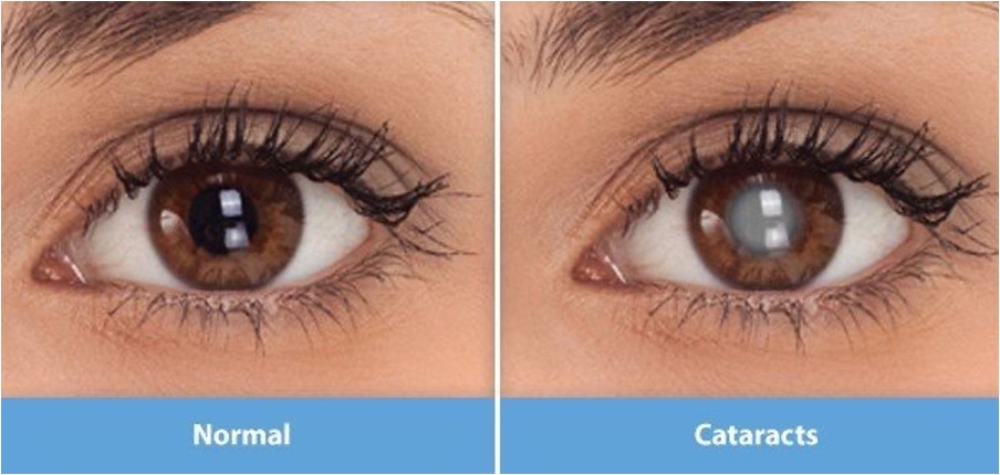
Cataract is a clouding of the natural lens in the eye which leads to a gradual drop of vision. Usually it develops gradually and can affect one or both eyes. Symptoms are faded colors, blurry vision, halos, and night vision problems.
The main cause of cataract is age, but other causes are related to systemic disease, certain systemic medications, eye trauma, radiation exposure and genetic disorders.
How do we treat cataract?
Nowadays treating cataract is very safe and effective. The procedure simply takes about 10 minutes, and the surgeon replaces your cloudy lens with a new artificial clear lens. The lens is positioned in the same place as your natural one and remains a permanent part of your eye.
The procedure is painless and it only requires topical (eye drops) anesthesia. Usually you get back to your regular life style within 2-3 days.
Treating cataract and avoids the need for distance and near glasses through one simple procedure.
What type of lenses can I have?
- Mono-focal IOLs (intra-ocular lenses):
It is the most common type of lenses where it replaces the cloudy lens and gives you crisp clear distance vision, but after the procedure you will need reading glasses for nearby objects. - Multi-focal IOLs (intra-ocular lenses):
This type of lenses has an advantage over the mono-focal IOLs as it is designed to give you clear vision at both distance and near, so after the procedure you will not need reading glasses.
Pediatric Ophthalmology
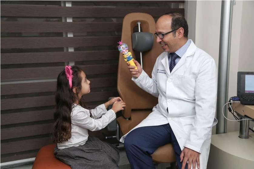
We put in your hands a multidisciplinary team of long specialized experts in the field of examining children's eyes. Our responsibility is to increase the awareness of the importance of examining children's eyes in a large segment of society because it is no less important than examining the child's growth scale, hearing and speech, levels of intelligence, dental and other health of the child. Some eye cases do not show any signs or symptoms, so the only way to be sure is to take your child for an eye test. In cases where there is no perceived problem and there is no significant family history of strabismus, lazy eye, or severe eye conditions in childhood, we recommend an annual eye exam from about 3 months to 9 years of age. Once these children reach the age of nine and over, we generally recommend an eye exam every two years unless your ophthalmologist advises otherwise.
Glaucoma: Importance of early diagnosis
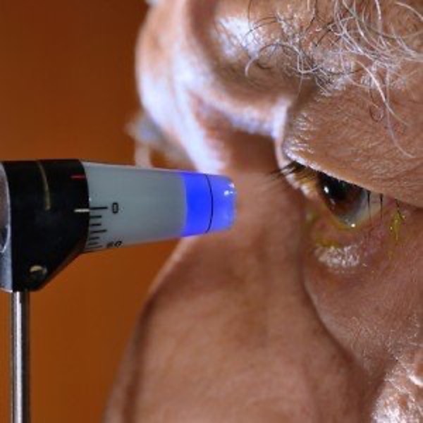
Glaucoma is high eye pressure, leading to damage to the optic nerve (the nerve that connects our eye to the brain). This can lead to permanent damage to the field of vision. In severe cases can cause tunnel vision and blindness. Even in less severe forms, glaucoma can interfere with independent living and can even affect our ability to drive.
Regular eye exams with your eye specialist, particularly if there is a family history of glaucoma, can detect the condition early. There are many sensitive tests that can help your ophthalmologist to follow up with your case including Visual Field tests and Optical Coherence Tomography scans. Both are done in clinic and only take a few minutes with no discomfort.
Once the diagnosis is confirmed, the key is to lower the eye pressure and preserve vision. At Abdali Hospital we offer the comprehensive treatment options under the supervision of an experienced Glaucoma Consultant. These include:
- Eye drops: There are a wide variety of eye drops which can be used to lower the eye pressure. They are usually taken once or twice a day but do need to be instilled regularly for the rest of your life.
- Selective Laser Trabeculoplasty: This is a simple and painless laser treatment that only takes a few minutes to perform and you can go home the same day. It can be done either to supplement the effect of eye drops or to replace them completely. At the time of your consultation, your consultant will discuss how this can help you.
- Laser Peripheral Iridotomy: In a particular type of glaucoma, narrow angle glaucoma, a very small channel is created in the iris (the coloured part of your eye) using a special laser. This can not only help lower the pressure but can also prevent future attacks of very high pressure (called acute angle closure glaucoma).
- Cyclodiode: If the eye pressure is not being controlled despite best efforts with all other treatment options, this laser can be performed to stop production of fluid in the eye (this is done by a part of the eye called the ciliary body).
- Surgery:
- Cataract Surgery: simply removing a developing cataract can not only lead to improvement in vision but can also lower the eye pressure.
- Trabeculectomy with antimetabolite injection: This involves creating a flap on the surface of the eye which allows fluid to drain out. A special anti-scarring medication is used to ensure success.
- Aqueous Shunt Devices: These devices comprise of a soft footplate connected to a plastic tube. The tube is inserted into the eye whilst the footplate is secured to the surface of the eye and drains fluid out. Performing either a trabeculectomy or aqueous shunt device insertion is reserved for cases that are not responding to other suitable treatments as listed above.
Early detection and prompt treatment of glaucoma is essential to preserve vision. An ongoing and strong support system surrounding you can make all the difference in living a fulfilling life despite glaucoma.
Corneal Topography
Corneal topography is performed for all patients asking for refractive surgery at their initial consultation. It is a comprehensive corneal scanner which provides data critical to planning any laser-surgical-treatment, giving us a proper assessment for refraction and helps in ruling out corneal diseases such as keratoconus (progressive thinning of the cornea) and the depth of any corneal scars. The corneal topography provides a truly accurate measurement of a patients cornea by taking 25,000 true elevation points and provides 3-dimensional analysis of the cornea. This includes a topographical assessment of both surfaces of the cornea, as well as the corneal thickness. By utilizing information from both the Belin / Ambrósio Enhanced Ectasia Display we analyze the data further to ensure regularity of the cornea and safety of the procedure.
Optical Coherence Tomography (OCT)
The Optical coherence tomography (OCT) is a safe, non-invasive medical imaging, that allows the light waves to penetrate the biological structures of the eye without harming any of the tissue as is free of ionizing lights or radiations. The OCT allows us to analyze several structures in the human eye. The most important are the optic nerve head, analysis of different retinal layers such as retinal nerve fiber layer thickness and ganglion cell complex, and macular volume. It’s a vital tool in glaucoma patients, patients with retinal diseases that affect the centre of the field of vision (such as in diabetic patients) as well as in patients with diseases that affect the optic nerve.